
Ronald A. Bergman, Ph.D., Adel K. Afifi, M.D., Paul M. Heidger,
Jr., Ph.D.
Peer Review Status: Externally Peer Reviewed
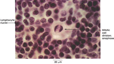
Human, Helly's fluid, H. & E., 612 x.
Lymphocyte nuclei: Densely staining, they fill most of the cell. The cytoplasm is not seen at this magnification but can be seen in Plate 53.
Mitotic cell division: Anaphase. Medium-sized and large lymphocytes are capable of multiplication by mitosis. Anaphase is a period in the continuous process of cell division in which the replicated chromosomes separate and are drawn toward the spindle poles prior to cytoplasmic division. For an illustration of the complete mitotic cycle and human chromosomes at high magnification see Plates 3 and 4.
Next Page | Previous Page | Section Top | Title Page
Please send us comments by filling out our Comment Form.
All contents copyright © 1995-2024 the Author(s) and Michael P. D'Alessandro, M.D. All rights reserved.
"Anatomy Atlases", the Anatomy Atlases logo, and "A digital library of anatomy information" are all Trademarks of Michael P. D'Alessandro, M.D.
Anatomy Atlases is funded in whole by Michael P. D'Alessandro, M.D. Advertising is not accepted.
Your personal information remains confidential and is not sold, leased, or given to any third party be they reliable or not.
The information contained in Anatomy Atlases is not a substitute for the medical care and advice of your physician. There may be variations in treatment that your physician may recommend based on individual facts and circumstances.
URL: http://www.anatomyatlases.org/