
Ronald A. Bergman, Ph.D., Adel K. Afifi, M.D., Paul M. Heidger,
Jr., Ph.D.
Peer Review Status: Externally Peer Reviewed
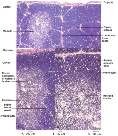
Human infant, 10% formalin, A. 35 x., B. 55 x., C. 87 x.
The primary function of the thymus is to produce immunocompetent T lymphocytes.
The three photomicrographs show the thymus at three levels of magnification.
A, The organ is highly lobated and is invested by a loose connective tissue capsule. From the capsule, connective tissue septa containing blood vessels penetrate the substance of the organ, forming lobes.
Lobes are composed of two distinctive regions, i.e., cortex and medulla. The outer cortical region stains intensely with basic dyes, such as hematoxylin, and appears almost uniformly blue. In addition, the cortex is punctuated by stellate reticular cells, which do not take a stain and appear as empty spaces. The cells cannot be seen at this magnification. The medulla, on the other hand, appears less blue (violet); it is less dense and contains eosinophilic structures (reticular fiber network and Hassall's corpuscles) and significantly fewer (not > 5 percent) lymphocytes.
Lymphoblasts found at the periphery of the cortex divide by mitosis producing small lymphocytes. The cortex contains small, medium, and large lymphocytes. The largest lymphocytes are about 9 µm, and have nuclei with abundant euchromatin and a strongly basophilic cytoplasm that contributes to the intense staining of the cortex.
Stellate reticular cells are part of the supporting framework of the organ and are not phagocytic cells. They are, however, believed to secrete thymic hormones, which promote the differentiation of T cells. Also importantly, the reticular cell processes effectively isolate developing lymphocytes and completely ensheath blood capillaries (the only type of blood vessel in the cortex) and thereby preclude the possibility of contaminating developing lymphocytes with antigens (blood-thymus barrier). Macrophages nevertheless are consistently found, however, in small numbers in the cortex.
The cortex produces massive numbers of lymphocytes, but the vast majority do not survive to leave the organ. Those that do leave the cortex do so via medullary postcapillary venules; these are immunocompetent T lymphocytes.
B, The thymic medulla contains primarily small, but fully mature, T lymphocytes. The T lymphocytes leave the medulla via venules and efferent lymphatic vessels. These lymphocytes will populate specific regions of other lymphoid organs such as lymph nodes (paracortical region) and spleen (periarterial sheaths of white pulp).
C, The thymic medulla characteristically contains Hassall's corpuscles (diagnostic feature), which vary in size from 25 to 200 µm in diameter. They consist of concentric layers of epithelial reticular cells, which are frequently degenerate, and may calcify.
Homologous and heterologous transplants are recognized as non-self foreign bodies. These tissues and organs are infiltrated by so-called graft rejection cells, which are T lymphocytes. These cells will disrupt the foreign tissue, resulting in rejection.
Next Page | Previous Page | Section Top | Title Page
Please send us comments by filling out our Comment Form.
All contents copyright © 1995-2024 the Author(s) and Michael P. D'Alessandro, M.D. All rights reserved.
"Anatomy Atlases", the Anatomy Atlases logo, and "A digital library of anatomy information" are all Trademarks of Michael P. D'Alessandro, M.D.
Anatomy Atlases is funded in whole by Michael P. D'Alessandro, M.D. Advertising is not accepted.
Your personal information remains confidential and is not sold, leased, or given to any third party be they reliable or not.
The information contained in Anatomy Atlases is not a substitute for the medical care and advice of your physician. There may be variations in treatment that your physician may recommend based on individual facts and circumstances.
URL: http://www.anatomyatlases.org/