
Plate 10.189 Esophagus-Stomach Junction
Ronald A. Bergman, Ph.D., Adel K. Afifi, M.D., Paul M. Heidger,
Jr., Ph.D.
Peer Review Status: Externally Peer Reviewed
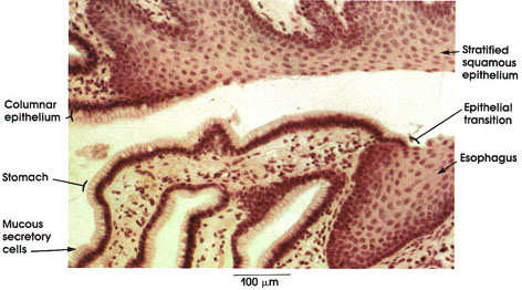
Dog, Helly's fluid, H. & E., 162 x.
Stratified squamous epithelium: Non-keratinized, it lines the esophagus. Indented by connective tissue papillae.
Epithelial transition: From the stratified squamous epithelium of the esophagus to the columnar epithelium of the stomach. Note that the transition is abrupt and that only the basal cells of the esophagus continue into the stomach.
Columnar epithelium: Tall simple columnar with basal nuclei. Continuous with the basal layers of esophagus epithelium. These cells secrete protective mucus constantly.
Next Page | Previous Page | Section Top | Title Page
Please send us comments by filling out our Comment Form.
All contents copyright © 1995-2024 the Author(s) and Michael P. D'Alessandro, M.D. All rights reserved.
"Anatomy Atlases", the Anatomy Atlases logo, and "A digital library of anatomy information" are all Trademarks of Michael P. D'Alessandro, M.D.
Anatomy Atlases is funded in whole by Michael P. D'Alessandro, M.D. Advertising is not accepted.
Your personal information remains confidential and is not sold, leased, or given to any third party be they reliable or not.
The information contained in Anatomy Atlases is not a substitute for the medical care and advice of your physician. There may be variations in treatment that your physician may recommend based on individual facts and circumstances.
URL: http://www.anatomyatlases.org/