
Plate 10.211 Submandibular Gland
Ronald A. Bergman, Ph.D., Adel K. Afifi, M.D., Paul M. Heidger,
Jr., Ph.D.
Peer Review Status: Externally Peer Reviewed
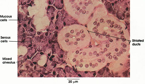
Human, Zenker's fluid, H. & E., 612 x
Mucous cells: Nuclei flattened and pushed to the basal part of the cell by secretory droplets. Purely mucous alveoli are not frequent in human submandibular gland.
Serous cells: Pyramidal in shape, darkly staining, with indistinct cell boundaries. Nuclei are more rounded and are pushed to the base of the cell by secretory droplets (zymogen granules) in some cells.
Mixed alveolus: Made up of serous and mucous cells. In mixed alveoli, serous cells cap mucous alveoli (so-called demilune) or line terminal portions of mucous alveoli.
Striated ducts: So-called because of prominent basal striations. These ducts are long and very conspicuous in sections of the submandibular gland. Lined by columnar cells with apically placed nuclei. Electron microscopy reveals the striations to be invaginations of the basal plasma membrane, with rows of elongated mitochondria in the pockets thus formed. The striated ducts play a role in secretion and absorption of salts and thereby modify the composition of the saliva produced by the secretory cells. The secretory product enters the oral cavity near the frenulum of the tongue. The submandibular gland produces about two thirds of the daily output of 1 liter of saliva. The saliva from this gland is a viscid solution containing mucin, salts, and the enzyme amylase.
Next Page | Previous Page | Section Top | Title Page
Please send us comments by filling out our Comment Form.
All contents copyright © 1995-2024 the Author(s) and Michael P. D'Alessandro, M.D. All rights reserved.
"Anatomy Atlases", the Anatomy Atlases logo, and "A digital library of anatomy information" are all Trademarks of Michael P. D'Alessandro, M.D.
Anatomy Atlases is funded in whole by Michael P. D'Alessandro, M.D. Advertising is not accepted.
Your personal information remains confidential and is not sold, leased, or given to any third party be they reliable or not.
The information contained in Anatomy Atlases is not a substitute for the medical care and advice of your physician. There may be variations in treatment that your physician may recommend based on individual facts and circumstances.
URL: http://www.anatomyatlases.org/