
Plate 10.217 Bile Canaliculi
Ronald A. Bergman, Ph.D., Adel K. Afifi, M.D., Paul M. Heidger,
Jr., Ph.D.
Peer Review Status: Externally Peer Reviewed
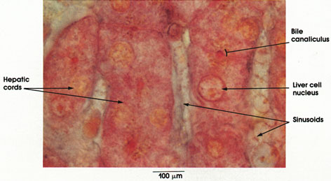
Human, Zenker's fluid, Mallory's stain, 1416 x.
Hepatic cords: See Plate 216.
Liver cell nucleus: Large and centrally placed.
Sinusoids: See Plate 216.
Bile canaliculi: Seen in cross section, these are channels between rows of cells within hepatic cords. Bile flows toward the periphery of the lobule to enter the system of bile ducts and gallbladder. Also see Plate 218.
Next Page | Previous Page | Section Top | Title Page
Please send us comments by filling out our Comment Form.
All contents copyright © 1995-2024 the Author(s) and Michael P. D'Alessandro, M.D. All rights reserved.
"Anatomy Atlases", the Anatomy Atlases logo, and "A digital library of anatomy information" are all Trademarks of Michael P. D'Alessandro, M.D.
Anatomy Atlases is funded in whole by Michael P. D'Alessandro, M.D. Advertising is not accepted.
Your personal information remains confidential and is not sold, leased, or given to any third party be they reliable or not.
The information contained in Anatomy Atlases is not a substitute for the medical care and advice of your physician. There may be variations in treatment that your physician may recommend based on individual facts and circumstances.
URL: http://www.anatomyatlases.org/