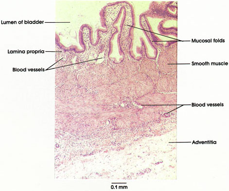

Plate 10.220 Gallbladder
Ronald A. Bergman, Ph.D., Adel K. Afifi, M.D., Paul M. Heidger,
Jr., Ph.D.
Peer Review Status: Externally Peer Reviewed

Human, 10% formalin, H. & E., 87 x.
The wall of the gallbladder consists of the following layers; (1) a mucosa composed of simple columnar epithelium, (2) a typical and unremarkable lamina propria, (3) a layer of smooth muscle, (4) an outer connective tissue layer, and (5) a typical serosa (where the free surface of the gallbladder contacts the peritoneum; otherwise, it is an adventitia).
Note the abundant folds of the mucosa, suggesting that the organ was empty and contracted when fixed for study, and the epithelium, which is a typical absorbing epithelium with microvilli but with the added ability to secrete small amounts of mucus.
The lamina propria contains loose connective tissue and small blood vessels.
The muscularis is relatively thin when the bladder is filled with bile but appears thick in this illustration because it is contracted. The smooth muscle is orientated circumferentially.
The main function of the gallbladder is to store and concentrate bile (by reabsorbing water) through what is called sodium pump activity.
Gallbladder contraction is induced by the hormone cholecystokinin, which is produced by enteroendocrine cells (so-called, I cells) located in the mucosal epithelium of the jejunurn and ileum. The secretion of cholecystokinin is initiated by dietary fats. A listing of these remarkable and uncommon cell types is given in the introductory material at the beginning of this section.
Next Page | Previous Page | Section Top | Title Page
Please send us comments by filling out our Comment Form.
All contents copyright © 1995-2024 the Author(s) and Michael P. D'Alessandro, M.D. All rights reserved.
"Anatomy Atlases", the Anatomy Atlases logo, and "A digital library of anatomy information" are all Trademarks of Michael P. D'Alessandro, M.D.
Anatomy Atlases is funded in whole by Michael P. D'Alessandro, M.D. Advertising is not accepted.
Your personal information remains confidential and is not sold, leased, or given to any third party be they reliable or not.
The information contained in Anatomy Atlases is not a substitute for the medical care and advice of your physician. There may be variations in treatment that your physician may recommend based on individual facts and circumstances.
URL: http://www.anatomyatlases.org/