
Plate 12.241 Ureter
Ronald A. Bergman, Ph.D., Adel K. Afifi, M.D., Paul M. Heidger,
Jr., Ph.D.
Peer Review Status: Externally Peer Reviewed
Human, 10% formalin, H. & E., 35 x.
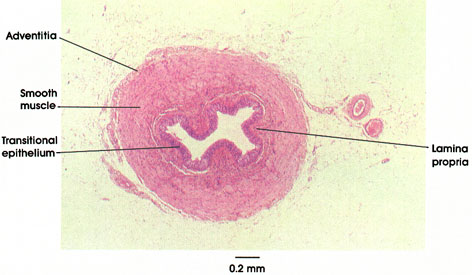
In cross section, the ureter typically exhibits a "festooned" appearance, reflecting contraction of the thick surrounding muscular layer and the consequent development of deep mucosal folds, consisting of the transitional epithelium and underlying lamina propria. There is no submucosa-the tissue of the lamina propria blends with the epimysial connective tissue of the muscularis. As in other urinary passages, the muscular layers are defined as inner longitudinal and outer circular (the converse of the gastrointestinal tract). However, these are not consistently clearly demarcated in histological section owing to incomplete separation of layers and intermingling of muscle fibers. The well-developed muscular layer generates the vigorous peristaltic wave, which propels urine from the renal pelvis to the urinary bladder. At the site of entry of the ureters into the bladder (the intramural portion of the ureter), the circular muscle layer disappears, and the ureters run an oblique course through the bladder wall. This anatomical arrangement, together with the closing of a fold of the bladder mucosa over the orifice of the ureter by hydrostatic backpressure of the bladder contents, provides an effective "physiological valve," which prevents backflow (reflux) of urine from the bladder to the ureter, and subsequently to the kidney pelvis. These anatomical features ensure against the kidney's being subject to hydrostatic pressure and tissue damage (hydronephrosis) upon voiding, and prevents reflux of bladder urine and transmission of infection to the upper urinary tract.
Next Page | Previous Page | Section Top | Title Page
Please send us comments by filling out our Comment Form.
All contents copyright © 1995-2024 the Author(s) and Michael P. D'Alessandro, M.D. All rights reserved.
"Anatomy Atlases", the Anatomy Atlases logo, and "A digital library of anatomy information" are all Trademarks of Michael P. D'Alessandro, M.D.
Anatomy Atlases is funded in whole by Michael P. D'Alessandro, M.D. Advertising is not accepted.
Your personal information remains confidential and is not sold, leased, or given to any third party be they reliable or not.
The information contained in Anatomy Atlases is not a substitute for the medical care and advice of your physician. There may be variations in treatment that your physician may recommend based on individual facts and circumstances.
URL: http://www.anatomyatlases.org/