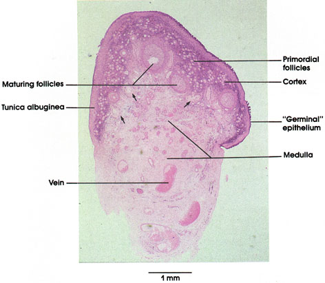

Plate 13.245 Ovary: Overview
Ronald A. Bergman, Ph.D., Adel K. Afifi, M.D., Paul M. Heidger,
Jr., Ph.D.
Peer Review Status: Externally Peer Reviewed

Monkey, glutaraldehyde, 1.5 µm, plastic section, H. & E., 47.5 x.
In this low magnification micrograph of primate ovary, the differentiation of the organ into cortical and medullary regions is seen. The highly vascular medulla is overlaid by a cortex in which various stages of follicular maturation may be identified. For example, the primordial follicles are most numerous and lie peripheral to growing follicles. Mature follicles (not shown here), which eventually ovulate, will have overgrown the entire width of the cortex. Follicles that have initiated growth but that have regressed are indicated by arrows and are termed atretic follicles.
The outer connective tissue investment, the tunica albuginea, is covered with a thin epithelium of peritoneal origin, the so-called germinal epithelium, named for its earlier, erroneously conceived role of seeding the ovary with germ cells.
Next Page | Previous Page | Section Top | Title Page
Please send us comments by filling out our Comment Form.
All contents copyright © 1995-2024 the Author(s) and Michael P. D'Alessandro, M.D. All rights reserved.
"Anatomy Atlases", the Anatomy Atlases logo, and "A digital library of anatomy information" are all Trademarks of Michael P. D'Alessandro, M.D.
Anatomy Atlases is funded in whole by Michael P. D'Alessandro, M.D. Advertising is not accepted.
Your personal information remains confidential and is not sold, leased, or given to any third party be they reliable or not.
The information contained in Anatomy Atlases is not a substitute for the medical care and advice of your physician. There may be variations in treatment that your physician may recommend based on individual facts and circumstances.
URL: http://www.anatomyatlases.org/