
Plate 13.252 Uterus
Ronald A. Bergman, Ph.D., Adel K. Afifi, M.D., Paul M. Heidger,
Jr., Ph.D.
Peer Review Status: Externally Peer Reviewed
Human, 10% formalin, H. & E., 50 x.
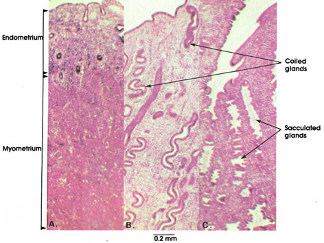
The uterine wall undergoes four phases during the menstrual cycle excluding menstruation. These are the (1) resurfacing, (2) proliferative, (3) secretory, and (4) ischemic phases.
The first three phases are shown in this plate. In A, the resurfacing phase, corresponding to days 5 and 6 of the cycle, is shown. During this stage, remnants of the glands in the basal zone of the mucosa proliferate and migrate to cover the raw surface of the endometrium denuded from its mucosa by menstrual flow. The thick myometrium is shown. This is a massive coat of smooth muscle fibers arranged in three concentric layers.
In B, the proliferative or follicular phase of the menstrual cycle, which lasts from day 7 to day 14 of the cycle, is shown. During this stage, the mucosal glands become longer and assume a curved or coiled configuration. The stroma between glands also increases by proliferation of connective tissue cells. The proliferative phase is induced by estrogen (see also Plate 249).
In C, the third or secretory phase, corresponding to days 15 to 27 of the menstrual cycle, is shown. This is also known as the progravid or luteal phase. During this stage, glands stop proliferating and begin to distend and secrete abundantly. in the middle region of the mucosa, saccular outpouchings of the glands are seen. The changes observed in this stage are induced by progesterone following estrogen priming.
Next Page | Previous Page | Section Top | Title Page
Please send us comments by filling out our Comment Form.
All contents copyright © 1995-2024 the Author(s) and Michael P. D'Alessandro, M.D. All rights reserved.
"Anatomy Atlases", the Anatomy Atlases logo, and "A digital library of anatomy information" are all Trademarks of Michael P. D'Alessandro, M.D.
Anatomy Atlases is funded in whole by Michael P. D'Alessandro, M.D. Advertising is not accepted.
Your personal information remains confidential and is not sold, leased, or given to any third party be they reliable or not.
The information contained in Anatomy Atlases is not a substitute for the medical care and advice of your physician. There may be variations in treatment that your physician may recommend based on individual facts and circumstances.
URL: http://www.anatomyatlases.org/