
Plate 13.254 Mammary Gland: Lactating
Ronald A. Bergman, Ph.D., Adel K. Afifi, M.D., Paul M. Heidger,
Jr., Ph.D.
Peer Review Status: Externally Peer Reviewed
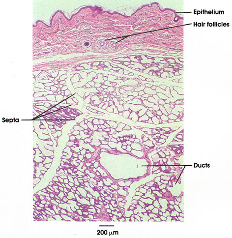
Monkey, 10% formalin, H. & E., 36 x.
A low power view of a section of actively lactating breast post parturn. The surface of the mammary gland is covered by a thin epithelium. The underlying dermis contains the follicles of fine surface hairs; the areola (not shown) is devoid of hairs. Alveoli are dilated and sacculated and contain a light- staining secretion product. With the degree of dilation shown by the secretory units, the smaller ducts are not distinguishable from the secretory units. Profiles of higher-order ducts are seen in the lower portion of the field.
Next Page | Previous Page | Section Top | Title Page
Please send us comments by filling out our Comment Form.
All contents copyright © 1995-2024 the Author(s) and Michael P. D'Alessandro, M.D. All rights reserved.
"Anatomy Atlases", the Anatomy Atlases logo, and "A digital library of anatomy information" are all Trademarks of Michael P. D'Alessandro, M.D.
Anatomy Atlases is funded in whole by Michael P. D'Alessandro, M.D. Advertising is not accepted.
Your personal information remains confidential and is not sold, leased, or given to any third party be they reliable or not.
The information contained in Anatomy Atlases is not a substitute for the medical care and advice of your physician. There may be variations in treatment that your physician may recommend based on individual facts and circumstances.
URL: http://www.anatomyatlases.org/