
Plate 14.262 Testis
Ronald A. Bergman, Ph.D., Adel K. Afifi, M.D., Paul M. Heidger,
Jr., Ph.D.
Peer Review Status: Externally Peer Reviewed
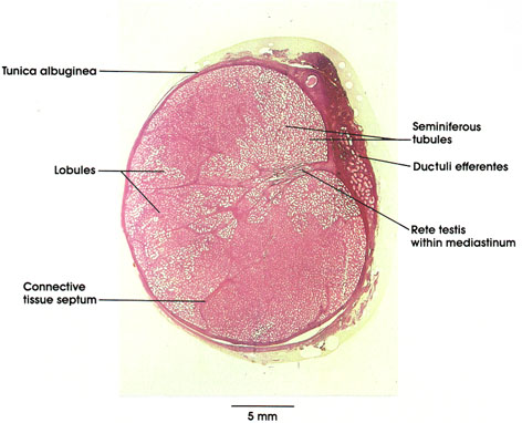
Human, 10% formalin, H. & E., 3.4 x.
The integrity of the delicate, tubular parenchyma of the male gonad is maintained by the robust connective tissue capsule of white fibrous connective tissue, the tunica albuginea, and the septa, which project interiorly, dividing the organ into lobules. The lobules contain the seminiferous tubules, lined with the specialized seminiferous epithelium, which gives rise to the male gametes, the spermatozoa. In addition, a posterior connective tissue mass (the mediastinum) projects to the interior of the organ and provides an avenue for the egress of fluid and spermatozoa from the seminiferous tubules. Irregular, epithelium-lined, anastomosing channels form a network, the rete, within the mediastinum. From these channels, the ductuli efferentes arise, pierce the tunica albuginea, and subsequently form, in man, the first portion of the epididymis.
Neither the seminiferous tubules nor the intratesticular duct system have significant muscular coats, or motile cilia by which to move the tubular contents. This underscores the role of fluid flux in moving the spermatozoa through the initial portion of the excurrent duct system. The ductuli efferentes are the sole portion of the male duct system that bears motile cilia on the lining epithelial cells.
The testis is invested on its anterior and lateral aspects by a serous envelope, the tunica vaginalis, which is a peritoneal vestige carried with the testis in its descent to the scrotum. The testis is cushioned by and glides within this serous envelope, relieving compression, to which the organ is exquisitely sensitive.
Next Page | Previous Page | Section Top | Title Page
Please send us comments by filling out our Comment Form.
All contents copyright © 1995-2024 the Author(s) and Michael P. D'Alessandro, M.D. All rights reserved.
"Anatomy Atlases", the Anatomy Atlases logo, and "A digital library of anatomy information" are all Trademarks of Michael P. D'Alessandro, M.D.
Anatomy Atlases is funded in whole by Michael P. D'Alessandro, M.D. Advertising is not accepted.
Your personal information remains confidential and is not sold, leased, or given to any third party be they reliable or not.
The information contained in Anatomy Atlases is not a substitute for the medical care and advice of your physician. There may be variations in treatment that your physician may recommend based on individual facts and circumstances.
URL: http://www.anatomyatlases.org/