
Plate 14.266 Testis
Ronald A. Bergman, Ph.D., Adel K. Afifi, M.D., Paul M. Heidger,
Jr., Ph.D.
Peer Review Status: Externally Peer Reviewed
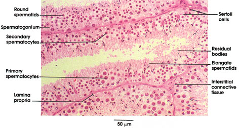
Monkey, glutaraldehyde, 1.5 µm plastic section, H. & E., 216 x.
Plastic-embedded, 1.5 µm, sections reveal to particular advantage the cell types constituting the seminiferous epithelium. The longitudinal section of a tubule in the center of the field contains many such representative cell types. Note the spermatogonia on the basement membrane. The largest of the germ cells, the primary spermatocytes with their characteristic meiotic prophase nuclei, occupy the region of the epithelium just apical to the basal layer of spermatogonia. The infrequently seen secondary spermatocytes are intermediate in size and placement within the epithelium between primary spermatocytes and spermatids.
Most apically situated within the epithelium are the spermatids, which possess either the smallest round nuclei of any cell of the epithelium or elongate, condensed nuclei, depending upon their stage of maturation toward mature spermatozoa.
The eosinophilic cytoplasmic remnants resulting from the differentiation of round spermatids to elongate spermatids are seen at the luminal surface and are termed residual bodies. Sustentacular cells, the Sertoli cells, phagocytize much of this residual cytoplasm. The nuclei of Sertoli cells are identified by their distinctive urn shape and prominent nucleolus. Note also how clusters of spermatids develop synchronously, enveloped within the apical extensions of Sertoli cell cytoplasm.
Adjacent seminiferous tubules share a common lamina propria; an area of interstitial connective tissue is shown where three adjacent tubules meet. Leydig cells are commonly found in such areas but are not present in this particular section.
Next Page | Previous Page | Section Top | Title Page
Please send us comments by filling out our Comment Form.
All contents copyright © 1995-2024 the Author(s) and Michael P. D'Alessandro, M.D. All rights reserved.
"Anatomy Atlases", the Anatomy Atlases logo, and "A digital library of anatomy information" are all Trademarks of Michael P. D'Alessandro, M.D.
Anatomy Atlases is funded in whole by Michael P. D'Alessandro, M.D. Advertising is not accepted.
Your personal information remains confidential and is not sold, leased, or given to any third party be they reliable or not.
The information contained in Anatomy Atlases is not a substitute for the medical care and advice of your physician. There may be variations in treatment that your physician may recommend based on individual facts and circumstances.
URL: http://www.anatomyatlases.org/