
Plate 15.285 Thyroid Gland
Ronald A. Bergman, Ph.D., Adel K. Afifi, M.D., Paul M. Heidger,
Jr., Ph.D.
Peer Review Status: Externally Peer Reviewed
Human, 10% formalin, H. & E., 162 x.
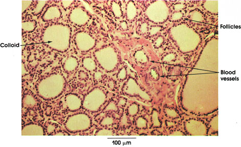
Follicles: Structural units of the thyroid gland. Note variations in shape (rounded or tubular) and size (0.05 to 0.5 mm in diameter). Close packing with a thin reticular network between adjacent follicles. Single layer of cells forms a hollow sphere. Nucleus centrally or basally placed.
Colloid: Found in the lumen of follicles. Chemical composition is a glycoprotein-iodine complex (thyroglobulin). Of the several iodinated compounds found in the gland the 3,5,3-triiodothyronine is hormonally the most active. The follicles release about 100 mg of hormone daily. Normal thyroid function is essential for the normal growth, development, and well-being of man and animals. Hypofunction of the thyroid in infants results in cretinism, characterized by dwarfism, mental deficiency, slow heart rate, muscular weakness, and gastrointestinal disturbances. Thyroid hormone given to infants at an early stage of cretinism can alleviate the symptoms. In adults, hypothyroidism results in muscular weakness, mental slowing, cold intolerance, and reflex and skin changes. When the hormone is produced in excess (hyperthyroidism), excessive appetite and thirst, weight loss, rapid respiration, sweating, muscular weakness, tremor, and an increase in heart rate (tachycardia) follow. Emotional disturbance and nervousness are also common symptoms.
Blood vessels: The thyroid is richly supplied with blood vessels, which are intimately associated with the follicles.
Next Page | Previous Page | Section Top | Title Page
Please send us comments by filling out our Comment Form.
All contents copyright © 1995-2024 the Author(s) and Michael P. D'Alessandro, M.D. All rights reserved.
"Anatomy Atlases", the Anatomy Atlases logo, and "A digital library of anatomy information" are all Trademarks of Michael P. D'Alessandro, M.D.
Anatomy Atlases is funded in whole by Michael P. D'Alessandro, M.D. Advertising is not accepted.
Your personal information remains confidential and is not sold, leased, or given to any third party be they reliable or not.
The information contained in Anatomy Atlases is not a substitute for the medical care and advice of your physician. There may be variations in treatment that your physician may recommend based on individual facts and circumstances.
URL: http://www.anatomyatlases.org/