
Plate 15.294 Adrenal Gland
Ronald A. Bergman, Ph.D., Adel K. Afifi, M.D., Paul M. Heidger,
Jr., Ph.D.
Peer Review Status: Externally Peer Reviewed
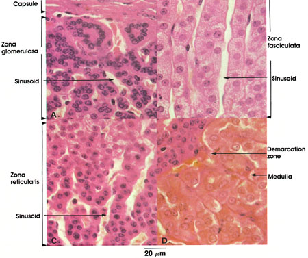
Rhesus monkey, Zenker's fluid, H. & E., 612 x.
This plate illustrates the different zones of the adrenal gland, from the capsule to the medulla.
Capsule: A tough fibroelastic covering with delicate trabeculae extending into the substance of the gland.
Zona glomerulosa: Outermost narrow zone of the adrenal cortex. Cells arranged in ovoid groups without a significant lumen. Component cells are columnar with spherical, deeply staining nuclei. Richly supplied with blood. The zona glomerulosa secretes hormones concerned primarily with mineral metabolism. The mineralocorticoids are deoxycorticosterone and aldosterone.
Sinusoid: Sinusoids arise from multiple arterioles in the capsule. Course between cell cords.
Zona fasciculata: The middle and broadest zone of the adrenal cortex. Cells are regularly arranged in parallel cords, one to two cells thick. Component cells are cuboidal, frequently containing two vesicular nuclei. Vacuoles seen in some cells represent dissolved lipid droplets. Cholesterol is chiefly present in this zone. Cell cords are surrounded by sinusoids. Secretes the glucocorticoids, cortisone and cortisol.
Zona reticularis: Innermost layer of the adrenal cortex. Cells are arranged in irregular cords, are smaller than those of the zona fasciculata, and stain darker. Nuclei stain deeply. Sinusoids separate cell cords. Secretes the same glucocorticoids as the zona fasciculata. These hormones participate in carbohydrate, protein, and fat metabolism.
Demarcation zone: Shows zone of transition from zona reticularis to medulla.
Medulla: Polyhedral cells arranged in anastomosing cords. Prominent nuclei. Contain chromaffin granules, precursors of epinephrine and norepinephrine. Richly supplied with blood.
The adrenal cortex is essential for life, and, through its hormones, is involved in numerous body functions. These activities include the maintenance of water and electrolyte balance, carbohydrate metabolism, and the normal functioning of connective tissue cells. Destruction or removal of the cortex results in Addison's* disease unless cortical hormones are given to the patient.
The adrenal medulla is not essential for life. The hormones of the adrenal medulla influence the metabolic rate and cardiovascular function and induce lipolysis and the release of fatty acids from adipose tissue, and opiate-like pepticles, including leu- and met-enkephalin. Medullary cells store their hormones in the form of granules.
During normal activity only a small amount of catecholamine is secreted, but this is greatly increased during stressful periods of intense emotional reaction. It is believed that epinephrine and norepinephrine are secreted by two different types of cells. The epinephrine secreting cells are primarily located around the distal ends of sinusoids draining the adrenal cortex, whereas norepinephrine-secreting cells are associated with capillaries of the direct arterial supply of the medulla. All adrenal medullary cells are innervated by cholinergic nerve terminals of preganglionic sympathetic neurons.
Chromaffin cells are also found in paraganglia, which are collections of catecholamine-secreting cells associated with autonomic ganglia. See Plate 295.
*Addison was a nineteenth-century English physician.
Next Page | Previous Page | Section Top | Title Page
Please send us comments by filling out our Comment Form.
All contents copyright © 1995-2024 the Author(s) and Michael P. D'Alessandro, M.D. All rights reserved.
"Anatomy Atlases", the Anatomy Atlases logo, and "A digital library of anatomy information" are all Trademarks of Michael P. D'Alessandro, M.D.
Anatomy Atlases is funded in whole by Michael P. D'Alessandro, M.D. Advertising is not accepted.
Your personal information remains confidential and is not sold, leased, or given to any third party be they reliable or not.
The information contained in Anatomy Atlases is not a substitute for the medical care and advice of your physician. There may be variations in treatment that your physician may recommend based on individual facts and circumstances.
URL: http://www.anatomyatlases.org/