
Plate 16.307 Retina
Ronald A. Bergman, Ph.D., Adel K. Afifi, M.D., Paul M. Heidger,
Jr., Ph.D.
Peer Review Status: Externally Peer Reviewed
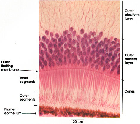
Rhesus monkey, Helly's fluid, H. & E., 612 x.
Outer plexiform layer: Contains axons of cone cells, dendrites of bipolar neurons, and processes of horizontal cells.
Outer nuclear layer: Nuclei of visual receptor cells. Note ovoid nuclei of the cones. Multiple rows of nuclei are characteristic of the fovea.
Outer limiting membrane: Sieve-like sheet. Region of junctional complexes between the outer ends of the tall supporting Müller cells and the adjoining photoreceptor cells. Not an actual membrane. The visual cells pass through perforations in this so-called membrane.
Cones: Light-sensitive end portions of cone cells. Respond to light of high intensity. Function is visual acuity and color perception. Compare cones in fovea with those seen in other regions of the retina (Plate 308).
Inner segments: Thicker proximal segments of the cones. Rich in mitochondria.
Outer segments: Slender distal segments of cones. Consist largely of stacked discs 0.014 µm thick. Contain iodopsin, an unstable, light-sensitive visual pigment, which chemically is composed of vitamin A, aldehyde conjugated to a specific protein (cone opsin).
Pigment epithelium: Single layer of pigmented cuboidal cells firmly bound to the choroid layer. Cells contain melanin pigment. The cytoplasmic processes of the pigment cells interdigitate with the outer segment of cones.
Next Page | Previous Page | Section Top | Title Page
Please send us comments by filling out our Comment Form.
All contents copyright © 1995-2024 the Author(s) and Michael P. D'Alessandro, M.D. All rights reserved.
"Anatomy Atlases", the Anatomy Atlases logo, and "A digital library of anatomy information" are all Trademarks of Michael P. D'Alessandro, M.D.
Anatomy Atlases is funded in whole by Michael P. D'Alessandro, M.D. Advertising is not accepted.
Your personal information remains confidential and is not sold, leased, or given to any third party be they reliable or not.
The information contained in Anatomy Atlases is not a substitute for the medical care and advice of your physician. There may be variations in treatment that your physician may recommend based on individual facts and circumstances.
URL: http://www.anatomyatlases.org/