
Plate 16.308 Eye
Ronald A. Bergman, Ph.D., Adel K. Afifi, M.D., Paul M. Heidger,
Jr., Ph.D.
Peer Review Status: Externally Peer Reviewed
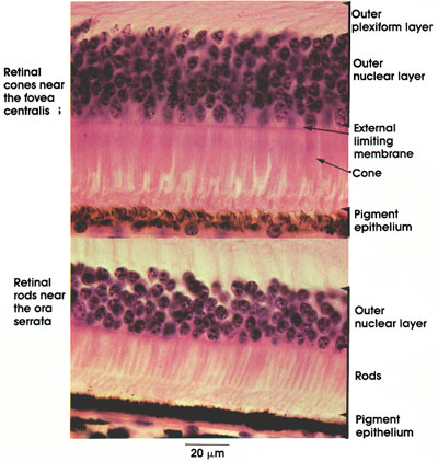
Rhesus monkey, 10% formalin, H. & E., 612 x.
This figure shows variation in retinal structure near the fovea centralis and at the periphery of the retina (ora serrata). In both sites, the layers of the retina are diminished. Absent are the ganglion cell layer, the inner plexiform layer, and the bipolar cell layer. The fovea constitutes the zone of greatest visual acuity. At the fovea, the photoreceptors are thin, slender elements (cones). They resemble rods more than cones. See Plate 307. The thinning of the retina at the fovea reduces to a minimum tissue through which light passes, and hence improves visual acuity. Note ovoid nuclei of cones in the outer nuclear layer. Multiple rows of these nuclei are characteristic of this region. Typical cones, which predominate here, function for sharp vision and color perception. It is estimated that there are 6 to 7 million cones in the retina.
Near the ora serrata, rods increase in number and in thickness, and become shorter. The cones decrease in number and also become shorter at the periphery of the retina. Rods number approximately 100 million and function for night vision and black and white discrimination. Note the multiple rows of rounded rod nuclei in the outer nuclear layer. The pigmented epithelium layer is similar in both fovea and ora serrata. It is firmly bound to the choroid layer and contains melanin pigment.
Next Page | Previous Page | Section Top | Title Page
Please send us comments by filling out our Comment Form.
All contents copyright © 1995-2024 the Author(s) and Michael P. D'Alessandro, M.D. All rights reserved.
"Anatomy Atlases", the Anatomy Atlases logo, and "A digital library of anatomy information" are all Trademarks of Michael P. D'Alessandro, M.D.
Anatomy Atlases is funded in whole by Michael P. D'Alessandro, M.D. Advertising is not accepted.
Your personal information remains confidential and is not sold, leased, or given to any third party be they reliable or not.
The information contained in Anatomy Atlases is not a substitute for the medical care and advice of your physician. There may be variations in treatment that your physician may recommend based on individual facts and circumstances.
URL: http://www.anatomyatlases.org/