
Plate 17.322 Spinal Cord: Sacral Region
Ronald A. Bergman, Ph.D., Adel K. Afifi, M.D., Paul M. Heidger,
Jr., Ph.D.
Peer Review Status: Externally Peer Reviewed
Human, Müller's fluid, Weigert's method with carmalum stain, 14 x.
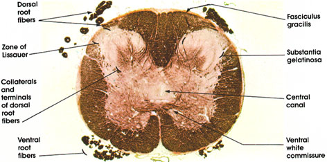
Dorsal root fibers: Bundles of heavily myelinated nerve fibers entering the spinal cord. Represent central processes of dorsal root ganglion neurons. Convey afferent impulses from peripheral organs to the spinal cord. Some of these fibers go directly to form tracts (fasciculus gracilis), others give collaterals or terminate on neurons in the spinal cord.
Zone of Lissauer*: Also known as fasciculus dorsolateralis. Composed of fine myelinated and non- myelinated fibers that carry pain, thermal, and light touch impulses or that interconnect different levels of the substantia gelatinosa.
Ventral root fibers: Axons of somatic, fusimotor, and visceral motor neurons in the anterior (ventral) and lateral gray columns. Heavily myelinated.
Fasciculus gracilis: Heavily myelinated ascending fiber system. Conveys kinesthetic sense and discriminative touch. Note the absence of the fasciculus cuneatus, which appears at spinal cord levels above T6.
Substantia gelatinosa: An expanded cell mass that forms the cap of the posterior gray horn of the spinal cord. Its size is related to that of the dorsal root. This area functions as an association region for incoming impulses. This region corresponds to lamina II of Rexed.
Central canal: Runs throughout the length of the cord. Partially obliterated in the adult.
Ventral white commissure: Bundle of myelinated fibers crossing from one side of the spinal cord to the other.
*Lissauer was a nineteenth-century German neurologist.
Next Page | Previous Page | Section Top | Title Page
Please send us comments by filling out our Comment Form.
All contents copyright © 1995-2024 the Author(s) and Michael P. D'Alessandro, M.D. All rights reserved.
"Anatomy Atlases", the Anatomy Atlases logo, and "A digital library of anatomy information" are all Trademarks of Michael P. D'Alessandro, M.D.
Anatomy Atlases is funded in whole by Michael P. D'Alessandro, M.D. Advertising is not accepted.
Your personal information remains confidential and is not sold, leased, or given to any third party be they reliable or not.
The information contained in Anatomy Atlases is not a substitute for the medical care and advice of your physician. There may be variations in treatment that your physician may recommend based on individual facts and circumstances.
URL: http://www.anatomyatlases.org/