
Plate 17.324 Medulla Oblongata
Ronald A. Bergman, Ph.D., Adel K. Afifi, M.D., Paul M. Heidger,
Jr., Ph.D.
Peer Review Status: Externally Peer Reviewed
Human, 10% formalin, Pal-Weigert and carmine stains, 11 x.
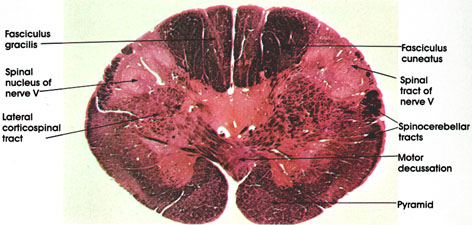
Fasciculus gracilis: Rostral continuation of the same tract seen at several spinal cord levels (see Plates 319 and 322). The lightly stained islands within the fasciculus represent neurons of the nucleus gracilis. This nucleus is larger at more rostral levels.
Fasciculus cuneatus: Rostral continuation of the same tract seen at spinal cord levels (Plate 319).
Spinal nucleus of nerve V: Functionally analogous to and structurally a continuation of the substantia gelatinosa seen at several spinal cord levels. In it terminate fibers of the descending (spinal) tract of cranial nerve V (trigeminal), which enters the neuraxis at a rostral level. The nucleus is primarily concerned with the perception of pain and thermal sense from the homolateral face.
Spinal tract of nerve V: Thinly myelinated fibers, hence less densely stained than the heavily myelinated fibers of the fasciculi gracilis and cuneatus or the spinocerebellar tracts. This tract is composed of descending trigeminal fibers and extends from the site of entry of the trigeminal nerve in the pons down to at least the second cervical spinal segment. Primarily concerned with pain and thermal sense. Synapse in the spinal nucleus of nerve V.
Spinocerebellar tracts: Heavily myelinated. Continuation of the same tracts seen at several spinal cord levels on their way to the cerebellum.
Motor decussation: Constitutes one of the most conspicuous features of sections at this level. Site of crossing of the pyramids to form the lateral corticospinal tracts. Approximately 75 to 90 per cent of descending pyramidal fibers cross at this level. The motor decussation forms the basis for voluntary motor control of one half of the body by the contralateral cerebral hemisphere.
Lateral corticospinal tract: Formed by decussation of the pyramidal tracts. Descends throughout the extent of the spinal cord (see Plate 325).
Pyramid: Heavily myelinated motor fiber system. Represents descending fibers from the cerebral cortex that pass through the internal capsule, cerebral peduncle, and pons before reaching the medullary pyramids. Fibers in the pyramid undergo partial crossing in the motor decussation to give rise to the lateral corticospinal tracts. It is estimated that, in man, about one million fibers are present in each pyramid.
Next Page | Previous Page | Section Top | Title Page
Please send us comments by filling out our Comment Form.
All contents copyright © 1995-2024 the Author(s) and Michael P. D'Alessandro, M.D. All rights reserved.
"Anatomy Atlases", the Anatomy Atlases logo, and "A digital library of anatomy information" are all Trademarks of Michael P. D'Alessandro, M.D.
Anatomy Atlases is funded in whole by Michael P. D'Alessandro, M.D. Advertising is not accepted.
Your personal information remains confidential and is not sold, leased, or given to any third party be they reliable or not.
The information contained in Anatomy Atlases is not a substitute for the medical care and advice of your physician. There may be variations in treatment that your physician may recommend based on individual facts and circumstances.
URL: http://www.anatomyatlases.org/