
Plate 17.326 Medula Oblongata
Ronald A. Bergman, Ph.D., Adel K. Afifi, M.D., Paul M. Heidger,
Jr., Ph.D.
Peer Review Status: Externally Peer Reviewed
Human, 10% formalin, Pal-Weigert, 7.5 x.
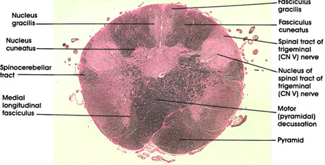
Nucleus gracilis: One of the dorsal column nuclei. Receives ascending fibers in the dorsal column (fasciculus gracilis) of the spinal cord entering below the level of the sixth thoracic spinal segment. Axons of neurons in this nucleus form the internal arcuate fiber system, cross in the midline (at more rostral levels), and form the medial lemniscus.
Fasciculus gracilis: Dorsal (posterior) column fibers capping the nucleus gracilis. Size is inversely proportional to that of nucleus gracilis, becoming smaller as more fibers terminate in the nucleus.
Nucleus cuneatus: One of the dorsal column nuclei. Located lateral to nucleus gracilis. Receives ascending fibers in the dorsal column of the spinal cord entering above the sixth thoracic spinal segment (fasciculus cuneatus). Axons of neurons in this nucleus, along with axons of the gracile neurons form the internal arcuate fiber system, which crosses in the midline to form the medial lemniscus.
Fasciculus cuneatus: Dorsal (posterior) column fibers capping the nucleus cuneatus. Size is inversely proportional to that of nucleus, becoming smaller as more fibers terminate in nucleus.
Spinal tract of trigerninal (CN V) nerve: Thinly myelinated fibers, hence less densely stained than the heavily myelinated fibers of the spinocerebellar tract. This tract is composed of descending trigeminal fibers and extends from the site of entry of the trigerninal nerve in the pons down to at least the second cervical spinal segment. Synapse in the nucleus of spinal tract of trigerninal nerve. Primarily concerned with pain and thermal sense from the homolateral face.
Nucleus of spinal tract of trigerninal (CN V) nerve: Functionally analogous to and structurally a continuation of the substantia gelatinosa seen at several spinal cord levels. In it terminate fibers of the descending (spinal) tract of cranial nerve V (trigeminal), which enters the neuraxis at a rostral level. The nucleus is primarily concerned with pain and thermal sense from the homolateral face.
Motor (pyramidal) decussation: Constitutes one of the most conspicuous features of sections at this level. Site of crossing of the pyramids to form the lateral corticospinal tracts. Approximately 75 to 90 percent of descending pyramidal fibers cross at this level. The motor decussation forms the basis for voluntary motor control of one half of the body by the contralateral cerebral hemisphere.
Pyramid: Corticospinal fibers prior to decussation.
Medial longitudinal fasciculus: Descending portion of a fiber system with ascending and descending components. Neurons of origin are from various brain stem nuclei with a major vestibular system component. Concerned with movement of neck and head in response to vestibular stimulation.
Spinocerebellar tract: Heavily myelinated. Continuation of the same tracts seen at several spinal cord levels. Destination is the cerebellum.
Next Page | Previous Page | Section Top | Title Page
Please send us comments by filling out our Comment Form.
All contents copyright © 1995-2024 the Author(s) and Michael P. D'Alessandro, M.D. All rights reserved.
"Anatomy Atlases", the Anatomy Atlases logo, and "A digital library of anatomy information" are all Trademarks of Michael P. D'Alessandro, M.D.
Anatomy Atlases is funded in whole by Michael P. D'Alessandro, M.D. Advertising is not accepted.
Your personal information remains confidential and is not sold, leased, or given to any third party be they reliable or not.
The information contained in Anatomy Atlases is not a substitute for the medical care and advice of your physician. There may be variations in treatment that your physician may recommend based on individual facts and circumstances.
URL: http://www.anatomyatlases.org/