
Plate 17.332 Medulla Oblongata
Ronald A. Bergman, Ph.D., Adel K. Afifi, M.D., Paul M. Heidger,
Jr., Ph.D.
Peer Review Status: Externally Peer Reviewed
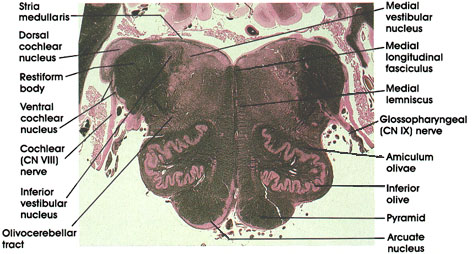
Human, 10% formalin, Pal-Weigert, 3.8 x.
Medial longitudinal fasciculus: Descending portion of a fiber system with ascending and descending components. Neurons of origin are from various brain stem nuclei, but with a major vestibular component. This system is concerned with eye and neck movements. The fibers in the descending component are destined to synapse on motor neurons in the cervical spinal cord that supply neck musculature.
Medial lemniscus: Continuation of the same system noted in caudal sections.
Glossopharyngeal (CN IX) nerve: A mixed nerve. Characteristically enters the medulla, inferior and medial to the restiform body.
Amiculurn olivae: A bundle of fibers surrounding the inferior olivary complex. Contains fibers that terminate on neurons of the olivary complex.
Inferior olive: Convoluted laminae of gray matter dorsal to the pyramid. Receives fibers from cortical and subcortical sites and projects fibers primarily to the contralateral but also to the ipsilateral cerebellum via the restiform body. Concerned with motor control.
Pyramid: Heavily myelinated motor fiber system. Contains descending fibers from the cerebral cortex that pass through the internal capsule, cerebral peduncle, and pons before reaching the pyramids. Fibers in the pyramid undergo partial crossing in the motor decussation caudal to this level.
Arcuate nucleus: Motor neurons ventral to the pyramid. Receives cortical input and projects to the cerebellum via the stria medullaris and restiform body. Homologous to pontine nuclei.
Olivocerebellar tract: Axons of neurons in the inferior olivary complex. Fibers arise from both olivary complexes but primarily from the contralateral complex. Destined for the cerebellum via inferior cerebellar peduncle (restiform body). Olivocerebellar fibers constitute the major component of the restiform body.
Cochlear (CN VIII) nerve: Central processes of bipolar neurons in the spiral ganglion. Enters the lateral surface of the pons lateral and dorsal to the restiform body. Projects upon the dorsal and ventral cochlear nuclei. Lesions in the cochlear nerve result in ipsilateral loss of hearing.
Ventral cochlear nucleus: Located ventral and lateral to the restiform body. Receives axons of the cochlear nerve originating in the upper turns of the cochlea.
Restiform body: Also known as inferior cerebellar peduncle. A compact bundle of nerve fibers connecting the medulla with the cerebellum. Tracts and fibers forming this bundle originate in the medulla and the spinal cord.
Dorsal cochlear nucleus: Characteristically located dorsal and lateral to the restiform body. Receives axons of the cochlear nerve originating in the lower turns of the cochlea.
Inferior vestibular nucleus: One of four vestibular nuclei. Characteristically located medial to the restiform body and traversed by myelinated bundles.
Stria medullaris: Axons of arcuate neurons. Courses in the floor of the fourth ventricle. Joins the restiform body to reach the cerebellum.
Medial vestibular nucleus: One of four vestibular nuclei. Characteristically located medial to the inferior vestibular nucleus in the floor of the fourth ventricle. Axons of neurons in this nucleus form the medial vestibulospinal tract.
Next Page | Previous Page | Section Top | Title Page
Please send us comments by filling out our Comment Form.
All contents copyright © 1995-2024 the Author(s) and Michael P. D'Alessandro, M.D. All rights reserved.
"Anatomy Atlases", the Anatomy Atlases logo, and "A digital library of anatomy information" are all Trademarks of Michael P. D'Alessandro, M.D.
Anatomy Atlases is funded in whole by Michael P. D'Alessandro, M.D. Advertising is not accepted.
Your personal information remains confidential and is not sold, leased, or given to any third party be they reliable or not.
The information contained in Anatomy Atlases is not a substitute for the medical care and advice of your physician. There may be variations in treatment that your physician may recommend based on individual facts and circumstances.
URL: http://www.anatomyatlases.org/