
Plate 17.336 Pons
Ronald A. Bergman, Ph.D., Adel K. Afifi, M.D., Paul M. Heidger,
Jr., Ph.D.
Peer Review Status: Externally Peer Reviewed
Human, 10% formalin, Pal-Weigert, 2.5 x.
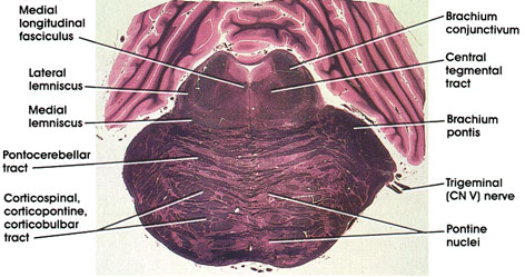
Brachium conjunctivum: Also known as superior cerebellar peduncle. Contains axons of deep cerebellar nuclei destined for the red nucleus and thalamus. Forms part of the lateral wall of the fourth ventricle.
Central tegmental tract: A compact fiber bundle located dorsal to the lateral part of the medial lemniscus. Carries fibers from midbrain tegmentum, red nucleus, and periaqueductal gray matter to the inferior olivary complex.
Brachium pontis: Also known as the middle cerebellar peduncle. Massive bundle of fibers connecting the basal portion of the pons with the cerebellum. Contains pontocerebellar fibers from the contralateral half of the pons. Some pontocerebellar fibers from the ipsilateral half of the pons are also contained in the brachium pontis.
Trigeminal (CN V) nerve: A mixed nerve with a larger sensory component (portio major) and a smaller motor component (portio minor).
Pontine nuclei: Located in the basal part of the pons. Continuous caudally with arcuate nuclei in the medulla oblongata. Receive corticofugal fibers and project (pontocerebellar tract) mainly to the contralateral cerebellum.
Corticospinal, corticopontine, corticobulbar tracts: A long descending fiber system sectioned transversely as it courses through the basal part of the pons.
Pontocerebellar tract: Axons of pontine nuclei destined for the cerebellum. Constitutes the major component of the middle cerebellar peduncle (brachium pontis).
Medial lemniscus: Continuation of the same fiber system noted in several more caudal levels. Note the change of orientation of this fiber bundle from a vertical orientation in the medulla to a horizontal orientation at this level.
Lateral lemniscus: Continuation of trapezoid body. Conveys auditory impulses.
Medial longitudinal fasciculus: The ascending component of this fasciculus. Contains fibers from the vestibular nuclei destined for the nuclei of extraocular movement. Lesions of the medial longitudinal fasciculus at this level will result in a characteristic clinical picture known as internuclear ophthalmoplegia.
Next Page | Previous Page | Section Top | Title Page
Please send us comments by filling out our Comment Form.
All contents copyright © 1995-2024 the Author(s) and Michael P. D'Alessandro, M.D. All rights reserved.
"Anatomy Atlases", the Anatomy Atlases logo, and "A digital library of anatomy information" are all Trademarks of Michael P. D'Alessandro, M.D.
Anatomy Atlases is funded in whole by Michael P. D'Alessandro, M.D. Advertising is not accepted.
Your personal information remains confidential and is not sold, leased, or given to any third party be they reliable or not.
The information contained in Anatomy Atlases is not a substitute for the medical care and advice of your physician. There may be variations in treatment that your physician may recommend based on individual facts and circumstances.
URL: http://www.anatomyatlases.org/