
Plate 17.339 Mesencephalon
Ronald A. Bergman, Ph.D., Adel K. Afifi, M.D., Paul M. Heidger,
Jr., Ph.D.
Peer Review Status: Externally Peer Reviewed
Human, 10% formalin, Pal-Weigert, 3.0 x.
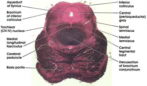
Inferior colliculus: Ovoid cellular mass belonging to the auditory system. Receives fibers from the lateral lemniscus and is reciprocally connected to the medial geniculate body.
Central (periaqueductal) gray: An area of gray matter surrounding the aqueduct of Sylvius. Contains scattered neurons, several nuclei, and some finely myelinated and unmyelinated fibers. Recent interest in this area has focused on its role in pain. The neuropeptide enkephalin has been identified in the central gray.
Spinal lemniscus (spinothalamic and spinotectal tracts): Continuation of the same fiber system seen at more caudal levels.
Medial lemniscus: Continuation of the same fiber system seen at more caudal levels.
Central tegmental tract: A compact fiber bundle located in the dorsal part of the mesencephalon dorsal to the decussation of brachiurn conjunctivurn. Carries fibers from the midbrain tegmentum, red nucleus, and periaqueductal gray matter to the inferior olivary complex. Note how the position of this tract changes in more caudal levels (see Plates 333 and 335, 336, 337).
Decussation of brachium conjunctivum: Massive outflow tract of the cerebellum seen crossing in the tegmenturn of the midbrain. Fibers project, after decussation, into the red nucleus and the ventral lateral nucleus of the thalamus. Lesion results in a disorder of coordinated movement.
Basis pontis: Basal part of pons. Contains pontine nuclei as well as corticospinal, corticobulbar, corticopontine, and pontocerebellar fibers.
Cerebral peduncle: Descending corticofugal fiber system. Lesion results in weakness (paresis) or paralysis of the contralateral half of the body, including the face.
Medial longitudinal fasciculus: The ascending component of this bundle. Connects vestibular nuclei with nuclei of extraocular movement (CN III, IV, VI).
Trochlear (CN IV) nucleus: Lies in the V-shaped ventral part of the central gray. Axons arch around the central gray, cross in the anterior medullary velum, and emerge from the dorsal aspect of the mesencephalon. Axons supply the superior oblique extraocular muscle.
Brachium of inferior colliculus: Also known as inferior quadrigeminal brachium. A bundle of nerve fibers from the lateral lemniscus and the inferior colliculus on their way to the medial geniculate body. This fiber bundle conveys auditory impulses from the midbrain to the thalamus.
Aqueduct of Sylvius: Named after the French anatomist Jacobus Sylvius (1478-1555). Connects the third and fourth ventricles.
Next Page | Previous Page | Section Top | Title Page
Please send us comments by filling out our Comment Form.
All contents copyright © 1995-2024 the Author(s) and Michael P. D'Alessandro, M.D. All rights reserved.
"Anatomy Atlases", the Anatomy Atlases logo, and "A digital library of anatomy information" are all Trademarks of Michael P. D'Alessandro, M.D.
Anatomy Atlases is funded in whole by Michael P. D'Alessandro, M.D. Advertising is not accepted.
Your personal information remains confidential and is not sold, leased, or given to any third party be they reliable or not.
The information contained in Anatomy Atlases is not a substitute for the medical care and advice of your physician. There may be variations in treatment that your physician may recommend based on individual facts and circumstances.
URL: http://www.anatomyatlases.org/