
Plate 17.340 Mesencephalon
Ronald A. Bergman, Ph.D., Adel K. Afifi, M.D., Paul M. Heidger,
Jr., Ph.D.
Peer Review Status: Externally Peer Reviewed
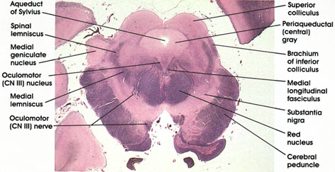
Human, 10% formalin, Pal-Weigert, 2.6 x.
Aqueduct of Sylvius: Connecting the third and fourth ventricles.
Periaqueductal (central) gray: Surrounds the aqueduct of Sylvius. Contains neurons related to both pain inhibition and stimulation.
Superior colliculus: Laminated cellular mass in the tecturn of the mesencephalon. Related to the visual system.
Brachium of inferior colliculus: Also known as inferior quadrigeminal brachium. Bundle of nerve fibers from the lateral lemniscus and the inferior colliculus to the medial geniculate body. Conveys auditory impulses to the thalamus.
Medial longitudinal fasciculus: Continuation of the same structure seen at more rostral and more caudal levels.
Substantia nigra: Largest nuclear mass in mesencephalon. Sandwiched between the medial lemniscus and the cerebral peduncle. A mass of pigmented cells (melanin) connected with basal ganglia and thalamus. Important in motor control. This area is invariably the site of pathological changes associated with Parkinson's disease. Parkinson was an eighteenth-century English physician.
Cerebral peduncle: Descending corticofugal fiber system. Lesions here result in weakness or paralysis of the contralateral half of the body, including the face.
Red nucleus: So-called because of a pinkish color in the fresh state owing to its high vascularity. This nucleus links the cerebellum, motor cortex, and spinal cord. Major input is from the brachium conjunctivurn and the cerebral cortex. Projects into the motor area of the cortex and spinal cord.
Oculomotor (CN III) nerve: Rootlets of the third cranial nerve seen coursing through the tegmenturn of the midbrain. Note relationship to red nucleus. Supplies the levator palpebrae superioris, the superior rectus, inferior rectus, medial rectus, inferior oblique, and constrictor pupillae muscles. Lesions of the oculomotor nerve result in ipsilateral paralysis of the eye muscles supplied by the nerve as well as a dilated, nonresponsive pupil.
Medial lemniscus: Continuation of the same tract seen at more caudal levels.
Medial geniculate nucleus: A thalamic nucleus concerned with audition. Receives auditory fibers from the inferior quadrigeminal brachiurn (brachiurn of inferior colliculus). Projects to the primary auditory cortex (transverse gyri of Heschl). Heschl was a nineteenth-century Austrian pathologist.
Spinal lemniscus: Continuation of the same structure seen at more caudal levels.
Oculomotor (CN III) nucleus: V-shaped collection of motor neurons located in a paramedian position, dorsal and medial to the medial longitudinal fasciculus. Axons form the oculomotor nerve. The oculomotor nucleus has two components: a somatic motor component, which supplies the extraocular (extrinsic) muscles, and a visceral, parasympathetic component (Edinger-Westphal nucleus), which supplies the intrinsic, constrictor pupillae muscle. Edinger was a nineteenth-century German anatomist and Westphal was a nineteenth-century German neurologist.
Next Page | Previous Page | Section Top | Title Page
Please send us comments by filling out our Comment Form.
All contents copyright © 1995-2024 the Author(s) and Michael P. D'Alessandro, M.D. All rights reserved.
"Anatomy Atlases", the Anatomy Atlases logo, and "A digital library of anatomy information" are all Trademarks of Michael P. D'Alessandro, M.D.
Anatomy Atlases is funded in whole by Michael P. D'Alessandro, M.D. Advertising is not accepted.
Your personal information remains confidential and is not sold, leased, or given to any third party be they reliable or not.
The information contained in Anatomy Atlases is not a substitute for the medical care and advice of your physician. There may be variations in treatment that your physician may recommend based on individual facts and circumstances.
URL: http://www.anatomyatlases.org/