
Plate 17.343 Diencephalon
Ronald A. Bergman, Ph.D., Adel K. Afifi, M.D., Paul M. Heidger,
Jr., Ph.D.
Peer Review Status: Externally Peer Reviewed
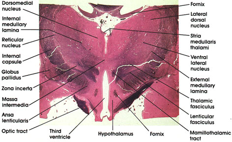
Human, 10% formalin, Pal-Weigert, 2.0 x.
Fornix: A paired C-shaped fiber tract connecting the hippocampus with several brain regions, including the mamillary body, anterior thalamic nucleus, septal nuclei, and cingulate gyrus. Seen in two locations in this figure, below the corpus callosurn and within the hypothalamus as it approaches its termination in the mamillary body.
Massa intermedia: Connects the two thalami across the third ventricle. Present in about 70 percent of human brains. Also referred to as the interthalamic adhesion.
Lateral dorsal (thalamic) nucleus: Belongs to the dorsal subgroup of the lateral thalamic nuclei. Has an association function.
Ventral lateral (thalamic) nucleus: Traversed by bundles of myelinated fibers. Located lateral to the internal medullary lamina. Receives fibers from the cerebellum via the brachium conjunctivum (dentatorubrothalamic fiber system). Has reciprocal connections with the primary motor cortex. Plays a role in motor control.
External medullary lamina: Defines the lateral boundary of the thalamus.
Thalamic fasciculus: A bundle of myelinated fibers destined for several thalamic nuclei from the basal ganglia and cerebellum.
Lenticular fasciculus: A bundle of myelinated fibers originating from the basal ganglia (globus pallidus) and destined for the thalamus via the thalamic fasciculus.
Mamillothalamic tract: Seen in cross section. Connects the mamillary body with the anterior thalamic nucleus.
Hypothalamus: Ventral to the thalamus. Forms part of the lateral wall of the third ventricle. The fornix separates medial and lateral zones of the hypothalamus.
Third ventricle: Between the two hypothalami and two thalami. Bisected by the massa intermedia.
Optic tract: Myelinated fiber bundle conveying visual impulses from the contralateral field of vision to the lateral geniculate nucleus.
Ansa lenticularis: Axons of neurons in globus pallidus coursing around the internal capsule. Projects to ventral anterior thalamic nucleus. Together with fibers from lenticular fasciculus forms the thalamic fasciculus.
Zona incerta: Sandwiched between the lenticular fasciculus and thalamic fasciculus. Rostral continuation of mesencephalic reticular formation. Continuous with the reticular nucleus of the thalamus.
Globus pallidus: Belongs to the basal ganglia along with the caudate nucleus and putamen. Located medial to the putamen and traversed by heavily myelinated fiber bundles. Comprises the principal efferent nucleus of the basal ganglia. Receives fibers from caudate, putamen, and subthalamic nuclei; and projects fibers to the thalamus (ventral anterior nucleus) and the subthalamic nucleus.
Internal capsule: The posterior limb of the internal capsule separates the basal ganglia and the thalamus. Massive cortical afferent and efferent bundle.
Internal medullary lamina: A band of myelinated fibers that separate the medial from the lateral group of thalamic nuclei. Contains fibers that interconnect different thalamic nuclei.
Reticular nucleus: A thalamic nucleus located between the internal capsule and external medullary lamina.
Dorsomedial (thalamic) nucleus: Belongs to the medial group of thalamic nuclei. Most highly developed in man. Located medial to the internal medullary lamina. Has reciprocal connections with the prefrontal cortex and hypothalamus. Receives input from other thalamic nuclei. Concerned with affective behavior and memory.
Stria medullaris thalami: Located clorsomedial to the thalamus. A component of the epithalamus. Connects the septal area and the habenular nuclei.
Next Page | Previous Page | Section Top | Title Page
Please send us comments by filling out our Comment Form.
All contents copyright © 1995-2024 the Author(s) and Michael P. D'Alessandro, M.D. All rights reserved.
"Anatomy Atlases", the Anatomy Atlases logo, and "A digital library of anatomy information" are all Trademarks of Michael P. D'Alessandro, M.D.
Anatomy Atlases is funded in whole by Michael P. D'Alessandro, M.D. Advertising is not accepted.
Your personal information remains confidential and is not sold, leased, or given to any third party be they reliable or not.
The information contained in Anatomy Atlases is not a substitute for the medical care and advice of your physician. There may be variations in treatment that your physician may recommend based on individual facts and circumstances.
URL: http://www.anatomyatlases.org/