
Atlas of Human Anatomy
Translated by: Ronald A. Bergman, PhD and Adel K. Afifi, MD, MS
Peer
Review Status: Internally Peer Reviewed
Magnified View (via Quicktime VR)
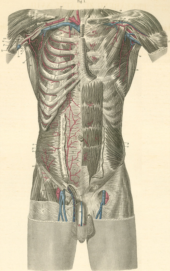
a) Clavicle.
b) First rib.
c) Second to tenth ribs.
d) Sternum (s. manubrium).
e) Xiphoid process of sternum.
f) m. Deltoideus.
g) m. Pectoralis major, humeral insertion.
g’) m. Pectoralis major, clavicular origin.
g’’) m. Pectoralis major, sternal origin.
h) m. Biceps brachii.
i) m. Coracobrachialis (on the right side the upper part of the muscle is divided,
to see the course of musculocutaneous nerve) (s. nervus perforans Casseri).
k) m. Pectoralis minor (right, only its insertion on the scapula is seen).
l) m. Subscapularis.
m) m. Latissimus dorsi.
n) m. Scalenus anterior.
o) m. Serratus anterior major (right, only the cut surface is seen).
p) mm. internal intercostales .
p’) m. First external intercostal.
q) m. Triangularis sterni.
r) m. Sternohyoideus.
s) m. Sternocleidomastoideus, sternal insertion.
s’) m. Sternocleidomastoideus, clavicular insertion.
t) m. Omohyoideus.
u) m. Trapezius.
v) m. Subclavius.
w) Costal pleural.
x) m. External abdominal oblique (both sides are cut).
y) m. Internal abdominal oblique (right side cut and left side is seen in situ).
z) m. Transversus abdominis.
a) m. Rectus abdominis.
b) Anterior part of the vagina (sheath)
of rectus abdominis (cut).
g) Umbilicus.
d) Linea alba.
e) m. Internal oblique (cut at the fleshy
end of the muscle).
z) Anterior part of the vagina of rectus
abdominis (cut).
h) Posterior part of the vagina of rectus
abdominis (also arcuate line s. linea semicircularis Douglasii).
k) Fascia transversa abdominis.
l) Inguinal ligament (Poupart’s ligament).
m) Abdominal annulus of the inguinal canal.
n) Spermatic cord (its middle is transversly
divided).
p) Suspensory ligament of the penis.
r) Femoral fascia lata.
s) Falciform process of fascia lata (the
outer end of the fossa ovalis is seen).
t) Floor of the fossa ovalis, formed by
the deep layer of fascia lata, which passes over the iliac fascia.
y) m. Sartorius.
f) m. iliopsoas.
w) m. pectineus.
Please send us comments by filling out our Comment Form.
All contents copyright © 1995-2024 the Author(s) and Michael P. D'Alessandro, M.D. All rights reserved.
"Anatomy Atlases", the Anatomy Atlases logo, and "A digital library of anatomy information" are all Trademarks of Michael P. D'Alessandro, M.D.
Anatomy Atlases is funded in whole by Michael P. D'Alessandro, M.D. Advertising is not accepted.
Your personal information remains confidential and is not sold, leased, or given to any third party be they reliable or not.
The information contained in Anatomy Atlases is not a substitute for the medical care and advice of your physician. There may be variations in treatment that your physician may recommend based on individual facts and circumstances.
URL: http://www.anatomyatlases.org/