
Atlas of Human Anatomy
Translated by: Ronald A. Bergman, PhD and Adel K. Afifi, MD, MS
Peer Review Status: Internally Peer Reviewed
Magnified View (via Quicktime VR)
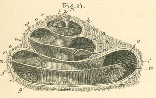
a) Cochlear base (s. basis).
b) Cupula (s. apex cochlea or helicotrema).
c) First turn of cochlear canal.
d) Second turn of cochlear canal.
e) Third (half) turn of cochlear canal.
f) Outer wall of cochlear canal.
g) Inner wall of cochlear canal.
h) Base of modiolus (s. basis der spindle).
i) Modiolus, spindle with hole and canal for a branch of the cochlear canal.
k) Apex modioli (location of helicotrema).
l) Fusion of modiolus with the cupula.
m) Bony spiral lamina.
n) Scala tympani (s. inferior).
o) Scala vestibuli (s. superior).
p) Terminus of cochlear canal (scyphus).
Please send us comments by filling out our Comment Form.
All contents copyright © 1995-2024 the Author(s) and Michael P. D'Alessandro, M.D. All rights reserved.
"Anatomy Atlases", the Anatomy Atlases logo, and "A digital library of anatomy information" are all Trademarks of Michael P. D'Alessandro, M.D.
Anatomy Atlases is funded in whole by Michael P. D'Alessandro, M.D. Advertising is not accepted.
Your personal information remains confidential and is not sold, leased, or given to any third party be they reliable or not.
The information contained in Anatomy Atlases is not a substitute for the medical care and advice of your physician. There may be variations in treatment that your physician may recommend based on individual facts and circumstances.
URL: http://www.anatomyatlases.org/