
Atlas of Human Anatomy in Cross Section: Section 1. Head and Neck
Ronald A. Bergman, Ph.D., Adel K. Afifi, M.D., Jean J. Jew, M.D., and Paul
C. Reimann, B.S.
Peer Review Status: Externally Peer Reviewed
|
Upper Left Quadrant |
Lower Left Quadrant |
Lower Right Quadrant |
Upper Right Quadrant |
|
1. Pericallosal br. of anterior cerebral a. |
12. Br. of middle cerebral a. |
19. Internal cerebral v. |
25. Claustrum |
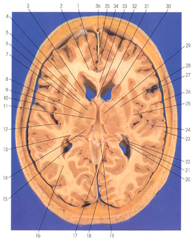
This is a section through the thalamus and basal ganglia. The two hemispheres are connected via the corpus callosum (2). Rostral and dorsal to the corpus callosum (2) on the medial surface of the hemisphere is the cingulate gyrus (34). In the interhemispheric fissure rostrally are pericallosal (1) and callosomarginal (35) branches of the anterior cerebral artery. Within the dural folds are seen two venous sinuses, a superficial superior sagittal sinus (36) and a deep straight sinus (17). The internal cerebral vein (19) is seen. The two frontal (anterior) horns of the lateral ventricle (31) are separated by the septum pellucidum (33). Ventral to the septum pellucidum (33) is the fornix (32). In the lateral wall of the frontal (anterior) horn of the lateral ventricle (31) is the head of the caudate nucleus (30). Ventral and lateral to the caudate nucleus (30) is the putamen (7). The caudate and putamen together form the striatum of the basal ganglia. The anterior limb of the internal capsule (4) separates the head of the caudate nucleus (30) from the putamen (7). The anterior limb of the internal capsule (4) is continuous with the genu (5) and the posterior limb (28) of the internal capsule. The internal capsule (4, 5, 28) contains corticofugal and corticopetal fibers. The posterior limb of the internal capsule (28) separates the putamen (7) from the thalamus. Within the thalamus, the following thalamic nuclei are seen: dorsomedial (29), ventral lateral (8), pulvinar (14), and anterior (3). The internal medullary lamina (6) separates the dorsomedial thalamic nucleus (29) from the ventral lateral thalamic nucleus (8). The internal medullary lamina (6) splits rostrally to enclose the anterior thalamic nucleus (3). The stria medullaris thalami (11) is seen coursing along the boundary of the thalamus. Lateral to the putamen is a slender band of gray matter, the claustrum (25). The choroid plexus (22) is seen within the trigone of the lateral ventricle (15). The hippocampus (20) bulges into the lateral ventricle (15). The alveus (21), representing axons of projection hippocampal neurons, is continuous with the fimbria of the fornix (13).
The central (rolandic) sulcus (27) separates the frontal and parietal lobes. The lateral (sylvian) fissure (10) separates the frontal, temporal, and parietal lobes. Branches of the middle cerebral artery (12) are seen within the lateral (sylvian) fissure (10). In the depth of the lateral fissure (10) is the insula (island of Reil) (9). On the lateral surface of the hemisphere, the parietal (26) and temporal (23) opercula cover the lateral (sylvian) fissure (10). The superior temporal gyrus (23) is continuous with the transverse gyrus of Heschl (24), the primary auditory cortex. In the posterior part of the hemisphere, the optic radiation (geniculocalcarine tract) (16) is seen coursing in the white matter to reach the striate (primary visual) cortex (18) on the medial surface of the occipital lobe.
Next Page | Previous Page | Section Top | Title Page
Please send us comments by filling out our Comment Form.
Anatomy Atlases is licensed under a Creative Commons Attribution-NonCommercial-ShareAlike 4.0 International License. 
"Anatomy Atlases", the Anatomy Atlases logo, and "A digital library of anatomy information" are all Trademarks of Michael P. D'Alessandro, M.D.
Anatomy Atlases is funded in whole by Michael P. D'Alessandro, M.D. Advertising is not accepted.
Your personal information remains confidential and is not sold, leased, or given to any third party be they reliable or not.
The information contained in Anatomy Atlases is not a substitute for the medical care and advice of your physician. There may be variations in treatment that your physician may recommend based on individual facts and circumstances.
URL: http://www.anatomyatlases.org/