
Plate 12.238 Kidney: Juxtaglomerular Cells
Ronald A. Bergman, Ph.D., Adel K. Afifi, M.D., Paul M. Heidger,
Jr., Ph.D.
Peer Review Status: Externally Peer Reviewed
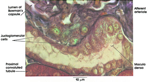
Rhesus monkey, Zenker's fluid, Mallory's stain, 1416 x.
Afferent arteriole: Terminal branch of the interlobular artery entering the glomerulus. The renal afferent arterioles are volume receptors and are sensitive to changes in perfusion (blood) pressure.
Juxtaglomerular cells: Granular variety of myoepithelioid cells in the wall of the afferent arteriole. Replace the typical smooth muscle cells of the tunica media of the artery. A decrease in afferent arterial volume secondary to low perfusion pressure results in the release of renin. Renin is an enzyme that is released into the blood and acts upon blood proteins to produce a potent vasoconstrictor, angiotensin, which can, under abnormal conditions, elevate blood pressure to dangerous levels. Hypertension of renal origin in humans can be cured by removal of the diseased or ischemic kidney. Renin also affects blood volume and osmolarity by initiating a chain of events leading to the release of the hormone aldosterone by the cells of the zona glomerulosa of the adrenal cortex. Aldosterone acts upon the renal tubules to enhance sodium reabsorption. A second system unrelated to the kidney (the hypothalamus of the brain and the posterior lobe of the pituitary, neurohypophysis), also regulates the volume and osmolarity of the extracellular fluid of the body. See Plates 116 and 237.
Macula densa: A group of specialized cells of the straight portion of the distal tubule, in contact with the afferent arteriole and contiguous with the juxtaglomerular cells. The cells are taller, thinner, and tightly packed compared to other distal tubule cells. These cells are functionally related to the juxtaglomerular cells, although their exact role is undefined. The macula densa marks the origin of the convoluted portion of the distal tubule.
Lumen of Bowman's capsule: Located between the parietal and visceral epithelial layers. Receives the ultrafiltrate of blood plasma circulating through the glomerular capillaries. The glomerular filtrate traverses the glomerular endothelium, the basal lamina (basement membrane), and the visceral epithelium to reach Bowman's space. Bowman's space is continuous with the lumen of the proximal convoluted tubule.
Proximal convoluted tubule: Deeply staining cuboidal cells surrounded by a thin basal lamina (basement membrane).
Next Page | Previous Page | Section Top | Title Page
Please send us comments by filling out our Comment Form.
Anatomy Atlases is licensed under a Creative Commons Attribution-NonCommercial-ShareAlike 4.0 International License. 
"Anatomy Atlases", the Anatomy Atlases logo, and "A digital library of anatomy information" are all Trademarks of Michael P. D'Alessandro, M.D.
Anatomy Atlases is funded in whole by Michael P. D'Alessandro, M.D. Advertising is not accepted.
Your personal information remains confidential and is not sold, leased, or given to any third party be they reliable or not.
The information contained in Anatomy Atlases is not a substitute for the medical care and advice of your physician. There may be variations in treatment that your physician may recommend based on individual facts and circumstances.
URL: http://www.anatomyatlases.org/