
Atlas of Human Anatomy in Cross Section: Section 1. Head and Neck
Ronald A. Bergman, Ph.D., Adel K. Afifi, M.D., Jean J. Jew, M.D., and Paul
C. Reimann, B.S.
Peer Review Status: Externally Peer Reviewed
|
Upper Left Quadrant |
Lower Left Quadrant |
Lower Right Quadrant |
Upper Right Quadrant |
|
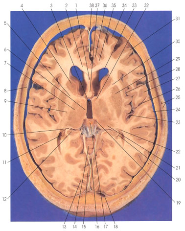
1. Septum pellucidum |
10. Fimbria of fornix |
16. Straight sinus |
24. Temporal operculum (superior temporal gyrus) |
This is a section (looking down) through the thalamus and basal ganglia. The two hemispheres are connected via the corpus callosum (2). Rostral and dorsal to the corpus callosum (2) on the medial surface of the hemisphere is the cingulate gyrus (36). The white matter core of the cingulate gyrus is the cingulum (35), an important long association fiber bundle. Within dural folds are seen three venous sinuses, the superior sagittal sinus (38), straight sinus (16), and the confluence of sinuses (18). The internal cerebral vein (19) is seen in close proximity to the pineal body (17). The two frontal (anterior) horns of the lateral ventricle (33) are seen separated by the septum pellucidum (1). Ventral to the septum pellucidum (1) are columns of fornix (37). In the lateral wall of the frontal (anterior) horn of the lateral ventricle (33) is the head of the caudate nucleus (4). Lateral and ventral to the head of the caudate nucleus (4) is the putamen (29). The caudate nucleus (4) and the putamen (29) are basal ganglia nuclei. They are separated by the anterior limb of the internal capsule (30). The anterior limb of the internal capsule (30) is continuous with the genu (5) and posterior limb (9) of the internal capsule. The internal capsule (5, 9, 30) carries corticofugal and corticopetal fibers. The posterior limb of the internal capsule (9) separates the putamen (29) from the thalamus. The following thalamic nuclei are seen: dorsomedial (6) in the lateral wall of the third ventricle (3), ventral lateral (7), pulvinar (21), and anterior (32). The temporal (inferior) horn of the lateral ventricle (11) is seen with the tail of the caudate nucleus (22) forming part of its roof and the hippocampus (12) protruding into it. The alveus (20), composed of axons of hippocampal projection neurons, is continuous with the fimbria of the fornix (10). The lateral (sylvian) fissure (26) is seen covered by frontal (27) and temporal (24) opercula. Branches of the middle cerebral artery (23) are seen within the lateral (sylvian) fissure (26). The insula (island of Reil) (28) is in the depth of the lateral (sylvian) fissure (26). The lingual gyrus (15) and the striate (primary visual) cortex (13) are seen on the medial surface of the occipital lobe. The superior (34) and middle (31) frontal gyri are seen in the frontal lobe. The superior surface of the vermis of the cerebellum (14) is seen in the midline. The middle meningeal artery (8) and the superficial temporal artery and vein (25) are seen.
Next Page | Previous Page | Section Top | Title Page
Please send us comments by filling out our Comment Form.
All contents copyright © 1995-2025 the Author(s) and Michael P. D'Alessandro, M.D. All rights reserved.
"Anatomy Atlases", the Anatomy Atlases logo, and "A digital library of anatomy information" are all Trademarks of Michael P. D'Alessandro, M.D.
Anatomy Atlases is funded in whole by Michael P. D'Alessandro, M.D. Advertising is not accepted.
Your personal information remains confidential and is not sold, leased, or given to any third party be they reliable or not.
The information contained in Anatomy Atlases is not a substitute for the medical care and advice of your physician. There may be variations in treatment that your physician may recommend based on individual facts and circumstances.
URL: http://www.anatomyatlases.org/