
Atlas of Human Anatomy in Cross Section: Section 1. Head and Neck
Ronald A. Bergman, Ph.D., Adel K. Afifi, M.D., Jean J. Jew, M.D., and Paul
C. Reimann, B.S.
Peer Review Status: Externally Peer Reviewed
|
Upper Left Quadrant |
Lower Left Quadrant |
Lower Right Quadrant |
Upper Right Quadrant |
|
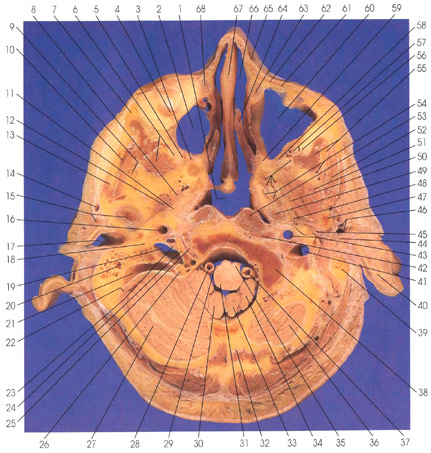
1. Nasopharynx |
17. External auditory meatus |
31. Medullary restiform body (inferior cerebellar peduncle) |
46. Superficial temporal a. and v. |
This section is similar to the one preceding, being its opposite face (looking down). This section passes through the nasal cavity (66), nasolacrimal ducts (63, 68), maxillary sinuses (2, 60), basioccipital bone (38), and the inferior part of the posterior cranial fossa, in which the cerebellar hemispheres (27, 36) can be seen. The section cuts the medulla oblongata (31, 35). Of particular interest is that cranial nerves 9 (glossopharyngeal) (25), 10 (vagus) (24), and 11 (spinal accessory) (23) are cut as they leave the cranial cavity via the jugular foramen and pass anterior to the jugular vein (19) medial to the internal carotid artery (16). Distally, they will lie between the carotid and the internal jugular vein. The superior vagal ganglion (26) is in the jugular foremen at the junction of the sigmoid sinus (21) and internal jugular vein (19).
The pterygopalatine ganglion (3) and the infraorbital artery (5) are identified.
The mandibular division of the trigeminal nerve is seen in the foremen ovale (8). The middle meningeal artery is seen in the foremen spinosum (12).
Next Page | Previous Page | Section Top | Title Page
Please send us comments by filling out our Comment Form.
All contents copyright © 1995-2024 the Author(s) and Michael P. D'Alessandro, M.D. All rights reserved.
"Anatomy Atlases", the Anatomy Atlases logo, and "A digital library of anatomy information" are all Trademarks of Michael P. D'Alessandro, M.D.
Anatomy Atlases is funded in whole by Michael P. D'Alessandro, M.D. Advertising is not accepted.
Your personal information remains confidential and is not sold, leased, or given to any third party be they reliable or not.
The information contained in Anatomy Atlases is not a substitute for the medical care and advice of your physician. There may be variations in treatment that your physician may recommend based on individual facts and circumstances.
URL: http://www.anatomyatlases.org/