
Atlas of Human Anatomy in Cross Section: Section 6. Pelvis, Perineum, Hip, and Upper Thigh
Ronald A. Bergman, Ph.D., Adel K. Afifi, M.D., Jean J. Jew, M.D., and Paul
C. Reimann, B.S.
Peer Review Status: Externally Peer Reviewed
|
Upper Left Quadrant |
Lower Left Quadrant |
Lower Right Quadrant |
Upper Right Quadrant |
|
1. Peritoneal cavity |
12. Nerve to iliacus m. |
20. Theca |
34. Femoral nerve |
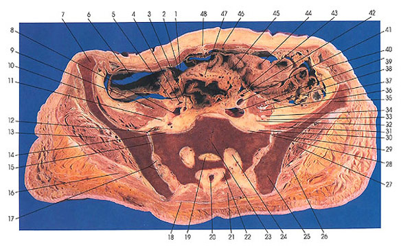
This section passes through the rectus abdominis muscles (46), ileum (44, 45), ascending (6) and descending (37) colon, sacroiliac joint (15, 28), and sacrum (23).
The following nerves can be identified: iliohypogastric and ilioinguinal (36), femoral (34), obturator (11, 33), fourth lumbar (32), nerve to iliacus (12, 30), first sacral (24), second sacral (22), and fifth lumbar (14). The theca (20) is still open at this level.
The left ureter crosses the common iliac artery (39), and the right ureter (3) crosses the common iliac artery at its point of bifurcation into external (4) and internal iliac (2) branches.
The anterior superior iliac spine of the ilium (7) is seen at this level.
The ureters (3, 43) lie medial to the ovarian arteries and veins (5, 43).
Next Page | Previous Page | Section Top | Title Page
Please send us comments by filling out our Comment Form.
All contents copyright © 1995-2024 the Author(s) and Michael P. D'Alessandro, M.D. All rights reserved.
"Anatomy Atlases", the Anatomy Atlases logo, and "A digital library of anatomy information" are all Trademarks of Michael P. D'Alessandro, M.D.
Anatomy Atlases is funded in whole by Michael P. D'Alessandro, M.D. Advertising is not accepted.
Your personal information remains confidential and is not sold, leased, or given to any third party be they reliable or not.
The information contained in Anatomy Atlases is not a substitute for the medical care and advice of your physician. There may be variations in treatment that your physician may recommend based on individual facts and circumstances.
URL: http://www.anatomyatlases.org/