
Atlas of Human Anatomy in Cross Section: Section 6. Pelvis, Perineum, Hip, and Upper Thigh
Ronald A. Bergman, Ph.D., Adel K. Afifi, M.D., Jean J. Jew, M.D., and Paul
C. Reimann, B.S.
Peer Review Status: Externally Peer Reviewed
|
Upper Left Quadrant |
Lower Left Quadrant |
Lower Right Quadrant |
Upper Right Quadrant |
|
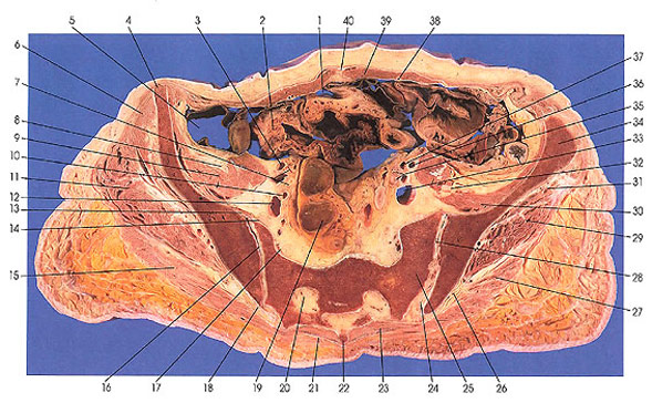
1. Peritoneal cavity |
13. Anterior sacroiliac ligament |
22. Second sacral vertebra spinous process |
31. Gluteus minimus m. |
This section passes through the linea alba (40) and rectus abdominis muscles (39), ascending (5), descending (33) and sigmoid (19) colon, sacroiliac joint (16, 28), and the second sacral vertebra (22).
The ovarian artery, vein, and nerve are still positioned anterior to the ureters (7, 35). This vascular and nervous unit is contained in a band or fold of the peritoneum that is called the suspensory ligament of the ovary. It may be traced craniad for some distance beyond the pelvic brim.
The gluteus medius (11, 29) on the outer surface of the iliac ala is now joined by the gluteus minimus (6, 31), which takes the deep position (third layer) from the inferoventral outer surface of the ala of the ilium. On the internal surface of the ala of the ilium the iliopsoas muscle (9) can be seen bilaterally.
Next Page | Previous Page | Section Top | Title Page
Please send us comments by filling out our Comment Form.
All contents copyright © 1995-2025 the Author(s) and Michael P. D'Alessandro, M.D. All rights reserved.
"Anatomy Atlases", the Anatomy Atlases logo, and "A digital library of anatomy information" are all Trademarks of Michael P. D'Alessandro, M.D.
Anatomy Atlases is funded in whole by Michael P. D'Alessandro, M.D. Advertising is not accepted.
Your personal information remains confidential and is not sold, leased, or given to any third party be they reliable or not.
The information contained in Anatomy Atlases is not a substitute for the medical care and advice of your physician. There may be variations in treatment that your physician may recommend based on individual facts and circumstances.
URL: http://www.anatomyatlases.org/