
Ronald A. Bergman, Ph.D., Adel K. Afifi, M.D., Paul M. Heidger,
Jr., Ph.D.
Peer Review Status: Externally Peer Reviewed

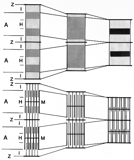
The upper figure, showing two sarcomeres, accounts for the usual light microscopic appearance (i.e., the staining densities) of sarcomeric cross striations in relaxed (left), contracting (middle), and fully contracted (right) skeletal and cardiac muscle fibers.
In the lower figure, also showing two sarcomeres, the comparable electron microscopic, ultrastructural configuration is shown. The appearance of cross striations or bands by both light and electron microscopy have their basis in the relative position and resulting density of the two major sets of myofilaments that constitute the sarcomere, that is, thin (actin) filaments, emanating from the Z line, and thick (myosin) filaments, held in hexagonal register at the M line, and their relative interdigitation in relaxation and in shortening (contraction).
Next Page | Previous Page | Section Top | Title Page
Please send us comments by filling out our Comment Form.
All contents copyright © 1995-2024 the Author(s) and Michael P. D'Alessandro, M.D. All rights reserved.
"Anatomy Atlases", the Anatomy Atlases logo, and "A digital library of anatomy information" are all Trademarks of Michael P. D'Alessandro, M.D.
Anatomy Atlases is funded in whole by Michael P. D'Alessandro, M.D. Advertising is not accepted.
Your personal information remains confidential and is not sold, leased, or given to any third party be they reliable or not.
The information contained in Anatomy Atlases is not a substitute for the medical care and advice of your physician. There may be variations in treatment that your physician may recommend based on individual facts and circumstances.
URL: http://www.anatomyatlases.org/