
Plate 10.206 Vermiform Appendix
Ronald A. Bergman, Ph.D., Adel K. Afifi, M.D., Paul M. Heidger,
Jr., Ph.D.
Peer Review Status: Externally Peer Reviewed
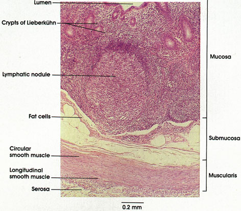
Human, 10% formalin, H. & E., 107 x.
The vermiform (worm-like) appendix extends from the cecum at the proximal end of the colon. The appendix has a structure similar to the colon except for the unusual longitudinal smooth muscle layer (taeniae coli, tapeworm of colon) of the latter, which terminates at the appendix and aids in its location when it is unusually placed.
The glands of the appendix are simple tubes, but they may fork. The glandular epithelium is rich in mucous cells and unicellular endocrine glands or enteroendocrine cells (EC). These cells are thought to secrete serotonin and substance P, which increase intestinal activity. The terms argyrophil, argentaffin, and enterochromaffin have been applied to these cells, which are rapidly gaining increased recognition for their importance to the function of the gastrointestinal system. See the introductory material at the beginning of this section for a listing of these cells. The surface epithelium is columnar with few mucous cells. Lymphatic nodules are abundant and intrusive and contain lymphatic nodules with germinal centers. See Plate 166, Lymphatic System. Both the nodules and massive lymphocytic infiltration are consipicuous histologic features that aid in the identification of this organ.
The lamina propria also characteristically contains fat cells.
The lumen of the organ is frequently filled with intestinal debris.
The biological significance of the appendix is unknown.
Next Page | Previous Page | Section Top | Title Page
Please send us comments by filling out our Comment Form.
All contents copyright © 1995-2024 the Author(s) and Michael P. D'Alessandro, M.D. All rights reserved.
"Anatomy Atlases", the Anatomy Atlases logo, and "A digital library of anatomy information" are all Trademarks of Michael P. D'Alessandro, M.D.
Anatomy Atlases is funded in whole by Michael P. D'Alessandro, M.D. Advertising is not accepted.
Your personal information remains confidential and is not sold, leased, or given to any third party be they reliable or not.
The information contained in Anatomy Atlases is not a substitute for the medical care and advice of your physician. There may be variations in treatment that your physician may recommend based on individual facts and circumstances.
URL: http://www.anatomyatlases.org/