
Polarization microscopy
Ronald A. Bergman, Ph.D., Adel K. Afifi, M.D., Paul M. Heidger,
Jr., Ph.D.
Peer Review Status: Externally Peer Reviewed

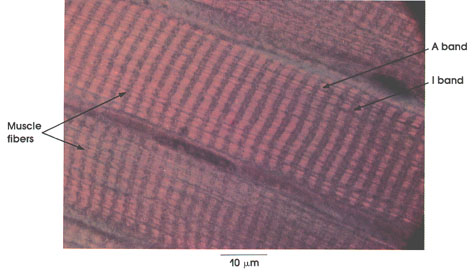
Human, Helly's fluid, H. & E., 1416 x.
The names given to the two major transverse striations of skeletal and cardiac muscle are derived from the studies of Brücke* (1858). With routine light microscopic techniques, alternating dark and light bands are seen within striated muscle fibers (Plates 66, 67 and 68). Polarization microscopy reverses the appearance of the dark band, which becomes bright, and the light band, which appears dark. The dark band of routine light microscopy, exhibiting birefringence with polarized light, is anisotropic and is called the A band. The light band of routine light microscopy is poorly refractile and relatively isotropic and is called the I band.
Muscle fibers: Showing cross striations formed by alternating segments of high and low refractive index resulting from their submicroscopic structure, which is revealed by electron microscopy.
A band: Anisotropic band.
I band: Isotropic band. Note the birefringence or anisotropy of the Z line in the center of the I band.
*Brücke was a nineteenth-century Viennese physiologist.
Next Page | Previous Page | Section Top | Title Page
Please send us comments by filling out our Comment Form.
All contents copyright © 1995-2024 the Author(s) and Michael P. D'Alessandro, M.D. All rights reserved.
"Anatomy Atlases", the Anatomy Atlases logo, and "A digital library of anatomy information" are all Trademarks of Michael P. D'Alessandro, M.D.
Anatomy Atlases is funded in whole by Michael P. D'Alessandro, M.D. Advertising is not accepted.
Your personal information remains confidential and is not sold, leased, or given to any third party be they reliable or not.
The information contained in Anatomy Atlases is not a substitute for the medical care and advice of your physician. There may be variations in treatment that your physician may recommend based on individual facts and circumstances.
URL: http://www.anatomyatlases.org/