
Plate 17.330 Medulla Oblongata
Ronald A. Bergman, Ph.D., Adel K. Afifi, M.D., Paul M. Heidger,
Jr., Ph.D.
Peer Review Status: Externally Peer Reviewed
Human, 10% formalin, Pal-Weigert and carmine stains, 7 x.
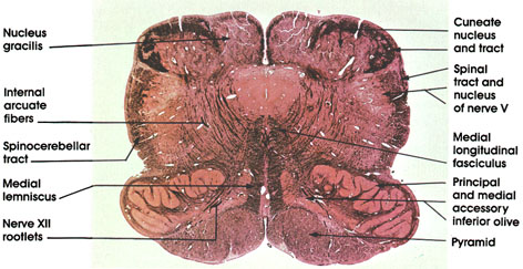
Nucleus gracilis: Fully developed at this level. Only a small remnant of the fasciculus gracilis caps the nucleus.
Cuneate nucleus and tract: Note that a definite portion of the fasciculus cuneatus caps the nucleus cuneatus as compared to that seen on the adjacent nucleus gracilis. The lightly stained island in the fasciculus cuneatus represents neurons of the accessory cuneate nucleus.
Spinal tract and nucleus of nerve V: Continuation of similar structures seen at more caudal levels (see Plates 324 and 328 ).
Medial longitudinal fasciculus: Note the change of position of the fasciculus in this figure as compared to a more caudal level (see Plate 328 ). This is a result of the formation of the medial lemniscus, which displaces the medial longitudinal fasciculus to a more dorsal location.
Principal and medial accessory inferior olive: This nuclear group distinguishes sections of the medulla at this level. The principal olive is the larger component with its hilum directed medially. The medial accessory olive is found along the border of the medial lemniscus. Inferior olive neurons give rise to olivocerebellar fibers that project into the cerebellum.
Pyramid: See the same structure at more caudal levels. Plates 324, 325, 326, 328, and 329.
Internal arcuate fibers: See also Plate 328, Axons of gracile and cuneate neurons.
Spinocerebellar tract: See also Plates 317, 324, 327, and 328. Continuation of the same tract seen in the spinal cord.
Medial lemniscus: Formed by the decussating internal arcuate fibers. Constitutes the second-order neurons of the posterior column pathways (fasciculi gracilis and cuneatus and their nuclei), conveying kinesthetic sense and discriminative touch to higher levels of the neuraxis.
Nerve XII rootlets: Hypoglossal cranial nerve. Note their characteristic location medial to the inferior olive and lateral to the pyramid. This proximity to the pyramid is the anatomical basis for the inferior or hypoglossal alternating hemiplegia resulting from lesions in this area. This syndrome (also known as medial medullary syndrome) consists of lower motor neuron paralysis of the ipsilateral half of the tongue and contralateral (upper motor neuron) hemiplegia. The hypoglossal nerve supplies all the intrinsic and extrinsic muscles of the tongue except the palatoglossus muscle.
Next Page | Previous Page | Section Top | Title Page
Please send us comments by filling out our Comment Form.
All contents copyright © 1995-2025 the Author(s) and Michael P. D'Alessandro, M.D. All rights reserved.
"Anatomy Atlases", the Anatomy Atlases logo, and "A digital library of anatomy information" are all Trademarks of Michael P. D'Alessandro, M.D.
Anatomy Atlases is funded in whole by Michael P. D'Alessandro, M.D. Advertising is not accepted.
Your personal information remains confidential and is not sold, leased, or given to any third party be they reliable or not.
The information contained in Anatomy Atlases is not a substitute for the medical care and advice of your physician. There may be variations in treatment that your physician may recommend based on individual facts and circumstances.
URL: http://www.anatomyatlases.org/